Full Body Skin Mapping
We are thrilled to bring the latest in skin care and mapping technology to our patients to provide an unparalleled experience in your health and treatment including fast and accurate skin mapping, to assist in proper analysis and treatment.
We are now offering DermoScan full body photography as well as VivaScope cellular skin imaging. Read on to learn more about what these imaging devices mean for helping to map, analyze, detect and diagnose skin changes in an innovative and minimally invasive way!
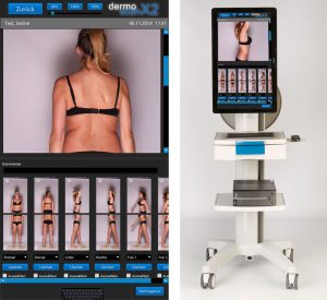
What is the DermoScan X2?
Developed for full-body skin mapping, the DermoScan X2 uses comprehensive technology to take full-body photographs. It is a complete system for identifying newly emergent pigmentation marks and diagnosing changes in existing lesions, using digital photo-documentation. The simple process relies on two, fixed-positioned cameras that provide a simultaneous photograph that allows for a 360 view of the body.
A complete preventative approach to addressing certain skin conditions requires regular monitoring of the whole body; changes in existing pigmentation marks or the appearance of new ones are important indicators for the early diagnosis of melanoma. TheDermoScan catches these sometimes imperceptible changes through past image comparison, analysis, and digital photo-documentation. The impressive technology can catch changes in size, shape and colour of existing or newly emergent lesions and depicts these alterations in an enlarged photograph.
DermoScan X2 is ideally suited to the documentation of all kinds of dermatoses, and is the perfect supplement to light microscopy.
It allows uniform, standardized pictures to be taken of the entire body, and the ability to compare pictures taken at different times (typically one year apart). The software allows us to see which moles are changing, and this is extremely helpful to us. On patients with multiple moles, it allows us to flag the changing moles, and view them side by side on the monitor in order to decide if the mole needs further examination or testing.
What is VivaScope?
VivaScope is a non-invasive system that performs an imaging “biopsy” to diagnose lesions without the traditionally invasive procedure. Using a low-powered laser the machine produces high-resolution images of the epidermis and the superficial collagen layers. This means that theVivaScope is able to look at living skin at the cellular level in real time.
Designed specifically to meet the stringent demands of clinicians and scientists investigating skin, VivaScope is well-suited for a wide variety of medical specialty areas where cellular level imaging of tissue can provide additional information necessary to support a clinical judgment. Additionally, theVivaScope tracks changes over time, meaning images can later be reviewed by the clinician to determine a diagnosis and course of treatment.
This non-invasive modality was the subject of Dr. Rubinstein’s fellowship at Harvard/MGH, where he did some of the earliest research on this modality. After many years of perfecting the technology, it is now available to us in the clinic. This device only takes a few minutes to acquire images, allows for instant diagnosis, is pain-free. The future of skin diagnosis is non-invasive, and we are proud to be one of the few dermatologists now offering this groundbreaking technology to our patients.
Call us or use our online scheduler to set up your appointment today!
*Individual results may vary; not a guarantee.
Schedule Now Patient Info Guide
Featured Treatment

RF Microneedling with Morpheus8
Morpheus8 combines microneedling with radiofrequency energy to provide deep skin tightening, wrinkle reduction, and facial and body contouring.* This FDA-cleared treatment is ideal for the lower face (jowls), and areas of the body, like arms or bra fat rolls. Morpheus8 is the first and only treatment to mold the fat deep in the dermal layers to “morph” and remodel skin tissue.
Learn MoreWhat's New
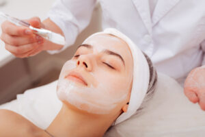
What Happens During a Professional Anti-Aging Firming Facial Treatment?
Skin naturally loses firmness and elasticity over time, and no amount of moisturizer or serum can fully replicate what a professional treatment can do.
Read MoreBack to Skin Basics